MouseVR
Development of MRI-compatible virtual reality goggles for fMRI in awake mice.
This project started through UNC's Summer Undergraduate Research Fellowship (SURF) and is being continued as the topic of my undergraduate honors thesis.
01 / THE QUESTION
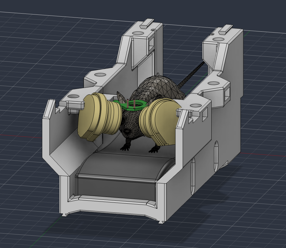
How can we deliver a virtual environment to an awake mouse during a whole-brain fMRI scan?
02 / WHY IT MATTERS
In traditional animal fMRI scans, the subject must be sedated to prevent head movements from corrupting image quality. This prevents the mouse from interacting meaningfully with a virtual environment and raises concerns about the sedated brain state being the “ground truth.”
However, the UNC Center for Animal MRI (CAMRI) has developed a novel technique for performing awake fMRI scans.1
With this technique, we hope to introduce a new experimental modality for animal fMRI researchers: virtual reality. When combined with CAMRI’s existing treadmill setup, many new and exciting experiments become possible, like traversing a maze.
03 / CONSTRAINTS
Size - everything must fit inside the scanner's small bore, together with the head-fixed mouse.
MRI Compatibility - magnetic components become dangerous when placed in the MRI, and even safe materials can disturb image quality by introducing unwanted artifacts.
04 / WHAT WE BUILT
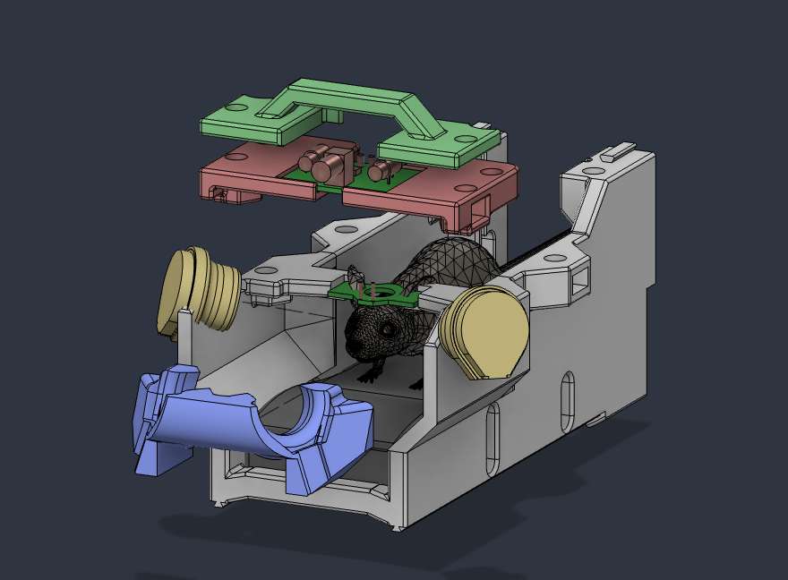
PLA 3D-printed enclosures, acrylic Fresnel lenses, and modified LED displays make up an MRI-compatible virtual reality system for mice.
05 / IMPACT
The goggles allow labs to perform experiments in controlled virtual environments while measuring the whole-brain activity of mice, creating a link between specific experiences and observed brain activity and behavior.
By revealing the links between experience, brain activity, learning, and emotion in mice, neuroscientists hope to discover foundational truths about these processes in human brains.